The Vagina

The vaginal canal passes upwards and backwards into the pelvis with the anterior and posterior walls in close con- tact along a line approximately parallel to the plane of the pelvic brim. When the woman stands upright, the vaginal canal points in an upward-backward direction and forms an angle of slightly more than 45 degrees with the uterus.
Function:
The vagina allows the escape of the menstrual fluids, receives the penis and the ejected sperm during sexual intercourse, and provides an exit for the fetus during birth.
Relations:
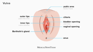
Knowledge of the relations of the vagina to other pelvic organs is essential for the accurate examination of the pregnant woman and the safe birth of the baby .Anterior to the vagina lie the bladder and the urethra, which are closely connected to the anterior vaginal wall.Posterior to the vagina lie the pouch of Douglas, the rectum and the perineal body, which separates the vagina from the anal canal.Laterally on the upper two-thirds are the pelvic fascia and the ureters, which pass beside the cervix; on either side of the lower third are the muscles of the pelvic floor.Superior to the vagina lies the uterus.Inferior to the vagina lies the external genitalia.
Structure:
The posterior wall of the vagina is 10 cm long, whereas the anterior wall is only 7.5 cm in length; this is because the cervix projects into its upper part at a right-angle.The upper end of the vagina is known as the vault. Where the cervix projects into it, the vault forms a circular recess that is described as four arches or fornices. The posterior fornix is the largest of these because the vagina is attached to the uterus at a higher level behind than in front. The anterior fornix lies in front of the cervix and the lateral fornices lie on either side.
Layers:
The vaginal wall is composed of three layers: mucosa, muscle and fascia. The mucosa is the most superficial layer and consists of stratified, squamous non-keratinized epithelium, thrown in transverse folds called ‘rugae. These allow the vaginal walls to stretch during intercourse and child- birth. Beneath the epithelium lies a layer of vascular connective tissue. The muscle layer is divided into a weak inner coat of circular fibres and a stronger outer coat of longitudinal fibres. Pelvic fascia surrounds the vagina and adjacent pelvic organs and allows for their independent expansion and contraction.There are no glands in the vagina; however, it is moistened by mucus from the cervix and a transudate that seeps out from the blood vessels of the vaginal wall.In spite of the alkaline mucus, the vaginal fluid is strongly acid (pH 4.5) owing to the presence of lactic acid formed by the action of Döderlein’s bacilli on glycogen found in the squamous epithelium of the lining. These lactobacilli are normal inhabitants of the vagina. The acid deters the growth of pathogenic bacteria.
Female Reproductive System
Blood Supply:
The blood supply comes from branches of the internal iliac artery and includes the vaginal artery and a descending branch of the uterine artery. The blood drains through corresponding veins.
Lymphatic Drainage:
Lymphatic drainage is via the inguinal, the internal iliac and the sacral glands.
Nerve Supply:
The nerve supply is derived from the pelvic plexus. The vaginal nerves follow the vaginal arteries to supply the vaginal walls and the erectile tissue of the vulva.