THE UTERUS

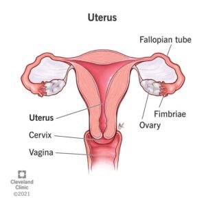
The uterus is a hollow, pear shaped muscular organ located in the true pelvis between the bladder and the rectum.The position of the uterus within the true pelvis is one of anteversion and anteflexion. Anteversion means that the uterus leans uterus forward and anteflexion means that it bends forwards upon itself. When the woman is standing, the uterus is in an almost horizontał position with the fundus resting on the bladder if the uterus is anteverted.
The term uterus is also applied to analogous structures in some non-mammalian animals.
In the human, the lower end of the uterus is a narrow part known as the isthmus that connects to the cervix, leading to the vagina. The upper end, the body of the uterus, is connected to the fallopian tubes, at the uterine horns, and the rounded part above the openings to the fallopian tubes is the fundus. The connection of the uterine cavity with a fallopian tube is called the uterotubal junction. The fertilized egg is carried to the uterus along the fallopian tube. It will have divided on its journey to form a blastocyst that will implant itself into the lining of the uterus the endometrium, where it will receive nutrients and develop into the embryo proper and later fetus for the duration of the pregnancy.
Function:
The main function of the uterus is to nourish the developing fetus prior to birth. It prepares for pregnancy each month and following pregnancy expels the products of conception.
Relations:
Knowledge of the relations of the uterus to other pelvic organs is desirable, particularly when giving women advice about bladder and bowel care during pregnancy and childbirth.
- Anterior to the uterus lie the uterovesical pouch and the bladder.
- Posterior to the uterus are the rectouterine pouch of Douglas and the rectum.
- Lateral to the uterus are the broad ligaments, the uterine tubes and the ovaries.
- Superior to the uterus lie the intestines.
- Inferior to the uterus is the vagina.
Supports:
The uterus is supported by the pelvic floor and maintained in position by several ligaments, of which those are the level of the cervix are the most important The transverse cervical ligaments fan out from the sides of the cervix to the side walls of the pelvis. They are sometimes known as the ‘cardinal ligaments’ or ‘Mackenrodt’s ligaments.
Structure:

The non-pregnant uterus is 7.5 cm long, 5 cm wide and 2.5 cm in depth, each wall being 1.25 cm thick . The cervix forms the lower-third of the uterus and measures 2.5 cm in each direction. The uterus consists of the following parts:
* The cornya are the upper outer angles of the uterus where the uterine tubes join.
* The fundus is the domed upper wall between the insertions of the uterine tubes.
* The body or corpus makes up the upper two-thirds of the uterus and is the greater part.
The cavity is a potential space between the anterior and posterior walls. It is triangular in shape, the base of the triangle being uppermost. . The isthmus is a narrow area between the cavity and the cervix, which is 7 mm long. It enlarges during pregnancy to form the lower uterine segment. . The cervix or neck protrudes into the vagina. The upper half, being above the vagina, is known as the supravaginal portion while the lower half is the infravaginal portion.
layers:

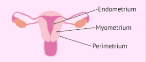
The uterus has three layers: the endometrium, the myometrium and the perimetrium, of which the myometrium, the middle muscle layer, is by far the thickest.
Blood Supply:
