THE PELVIS

Knowledge of anatomy of a normal female pelvis is key to midwifery and obstetric practice, as one of the ways to estimate a woman’s progress in labour is by assessing the relationship of the fetus to certain bony landmarks of the pelvis. Understanding the normal pelvic anatomy helps to detect deviations from normal and facilitate appropriate care.
The Pelvic Girdle:
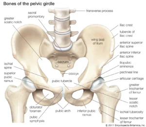
The pelvic girdle is a basin-shaped cavity and consists of two innominate bones (hip bones), the sacrum and the coccyx. It is virtually incapable of independent movement except during childbirth, as it provides the skeletal framework of the birth canal. It contains and protects the bladder, rectum and internal reproductive organs.
In addition, it provides an attachment for trunk and limb muscles. Some women experience pelvic girdle pain in pregnancy and need referral to a physiotherapist .
Innominate Bones:
Each innominate bone or hip bone is made up of three bones that have fused together: the ilium, the ischium and the pubis.
On its lateral aspect is a large, cup-shaped acetabulum articulating with the femoral Et head, which is composed of the three fused bones in d the following proportions: two-fifths ilium, two-fifths ischium and one-fifth pubis Anteroinferior to this is the large oval or triangular obturator for a- men. The bone is articulated with its fellow to form the pelvic girdle.
The ilium has an upper and lower part. The smaller lower part forms part of the acetabulum and the upper part is the large flared-out part. When the hand is placed on the hip, it rests on the iliac crest, which is the upper border. A bony prominence felt in front of the iliac crest is known as the anterior superior iliac spine.
A short distance below it is the anterior inferior iliac spine. There are two similar points at the other end of the iliac crest, namely the posterior superior and the posterior inferior iliac spines. The internal concave anterior surface of the ilium is known as the iliac fossa.
The ischium Is the inferoposterior part of the innom– inate bone and consists of a body and a ramus. Above, it forms part of the acetabulum. Below its ramus, it ascends anteromedially at an acute angle to meet the descending pubic ramus and complete the obturator foramen.
It has a large prominence known as the ischial tuberosity on which the body rests when sitting. Behind and a little above the tuberosity is an inward projection, the ischial spine. This is an important landmark in midwifery and obstetric practice, as in labour, the station of the fetal head is estimated In relation to the ischial spines allow- ing assessment of progress of labour.
The pubis forms the anterior part. It has a body and two oar-like projections, the superior ramus and the infe– rior ramus. The two pubic bones meet at the symphysis pubis and the two inferior rami form the pubic arch, merging into a similar ramus on the ischium. The space enclosed by the body of the pubic bone, the rami and the ischium is called the ‘obturator foramen.
The Sacrum:
The sacrum is a wedge-shaped bone consisting of five fused vertebrae, and forms the posterior wall of the pelviccavity as it is wedged between the Innominate bones. The caudal apex articulates with the coccyx and the upper border of the first sacral vertebra (sacral promontory) articulates with the first lumbar vertebra. The anterior surface of the sacrum is concave and is referred to as the hollow of the sacrum.
Laterally, the sacrum extends into a wing or ala. Four pairs of holes or foramina pierce the sacrum and, through these, nerves from the cauda equina emerge to innervate the pelvic organs. The posterior sur- face is roughened to receive attachments of muscles.
The Coccyx:
The coccyx is a vestigial tail. It consists of four fused vertebrae, forming a small triangular bone, which articulates with the fifth sacral segment.