The Pelvis in Relation to Pregnancy and Childbirth:
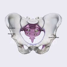
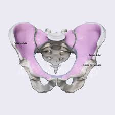
The term ‘pelvis’ is applied to the skeletal ring formed by the innominate bones and the sacrum, the cavity within and even the entire region where the trunk and the lower limbs meet. The pelvis is divided by an oblique plane, which passes through the prominence of the sacrum, the arcuate line (the smooth rounded border on the internal surface of the ilium), the pectineal line (a ridge on the superior ramus of the pubic bone) and the upper margin of the symphysis pubis, into the true and the false pelvis.
The True Pelvis:

The true (or lesser) pelvis is bounded in front and below by the pubic symphysis and the superior rami of the pubis; above and behind, by the sacrum and coccyx; and laterally, by a broad, smooth, quadrangular area of bone, corresponding to the inner surfaces of the body and superior ramus of the ischium, and the part of the ilium below the arcuate line.
The true pelvis is the bony canal through which the fetus must pass during birth. It is divided into a brim, a cavity and an outlet.
The Pelvic Brim:
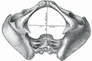
The superior circumference forms the brim of the true pelvis, the included space being called ‘the inlet. The brim is round except where the sacral promontory projects into it.
Midwives need to be familiar with the fixed points on the pelvic brim that are known as its landmarks, Commencing posteriorly, these are:
(1) Sacral promontory
(2) Sacral ala or wing
(3) Sacroiliac joint
(4) iliopectineal line, which is the edge formed at the inward aspect of the ilium
(5) iliopectineal eminence, which is a roughened area formed where the superior ramus of the pubic bone meets the ilium
(6) superior ramus of the pubic bone
(7) upper inner border of the body of the pubic bone
(8) upper inner border of the symphysis pubis.
The Pelvic Cavity

The cavity of the true pelvis extends from the brim superiorly to the outlet inferiorly. The anterior wall is formed by the pubic bones and symphysis pubis and its depth is 4 cm. The posterior wall is formed by the curve of the sacrum, which is 12 cm in length. Because there is such a difference in these measurements, the cavity forms a curved canal. With the woman upright, the upper portion of the pelvic canal is directed downward and backward, and its lower course curves and becomes directed downward and forward. Its lateral walls are the sides of the pelvis, which are mainly covered by the obturator internus muscle.
The cavity contains the pelvic colon, rectum, bladder and some of the reproductive organs. The rectum is placed posteriorly, in the curve of the sacrum and coccyx; the bladder is anterior behind the symphysis pubis
The Pelvic Outlet

The lower circumference of the true pelvis is very irregular; the space enclosed by it is called the outlet. Two outlets are described: the anatomical and the obstetrical.
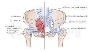
THE ANATOMICAL OUTLET The anatomical outlet is formed by the lower borders of each of the bones together with the sacrotuberous ligament.
THE OBSTETRICAL OUTLET
The obstetrical outlet is of greater practical significance because it includes the narrow pelvic strait through which the fetus must pass. The narrow pelvic strait lies between the sacrococcygeal joint, the two ischial spines and the lower border of the symphysis pubis. The obstetrical outlet is the space between the narrow pelvic strait and the anatomical outlet. This outlet is diamond-shaped.
The False Pelvis

It is bounded posteriorly by the lumbar vertebrae and laterally by the iliac fossae, and in front by the lower portion of the anterior abdominal wall. The false pelvis varies considerably in size according to the flare of the iliac bones. However, the false pelvis has no significance point in midwifery.