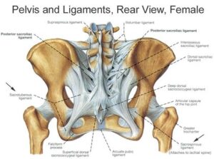
Pelvic Joints
There are four pelvic joints: one symphysis pubis, two sacroiliac joints and one sacrococcygeal joint.
• The symphysis pubis is the midline cartilaginous joint uniting the rami of the left and right pubic bones.
• The sacroiliac joints are strong, weight-bearing synovial joints with irregular elevations and depressions that produce interlocking of the bones. They join the sacrum to the ilium and as a result connect the spine to the pelvis. The joints allow a limited backward and forward movement of the tip and promontory
Of the sacrum, sometimes known as ‘nodding’ of the sacrum.
• The sacrococcygeal joint is formed where the base of the coccyx articulates with the tip of the sacrum. It permits the coccyx to be deflected backwards during the birth of the fetal head.
Pelvic Ligaments
The pelvic joints are held together by very strong ligaments that are designed not to allow
movement. However, during pregnancy the hormone relaxin gradually loosens all the
pelvic ligaments allowing slight pelvic movement thereby providing more room for the fetal
head as it passes through the pelvis. A widening of 2-3 mm at the symphysis pubis during
pregnancy above the normal gap of 4-5 mm is normal but if it widens significantly, the
degree of movement permitted may give rise to pain on walking.
The ligaments connecting the bones of the pelvis with each other can be divided into four
groups:
those connecting the sacrum and ilium – the sacroiliac ligaments
those passing between the sacrum and ischium – the sacrotuberous ligaments and the
sacrospinous ligaments those uniting the sacrum and coccyx – the sacrococCygeal ligaments
The Pelvis in Relation to Pregnancy and Childbirth
The term ‘pelvis’ is applied to the skeletal ring formed by the innominate bones and the
sacrum, the cavity within and even the entire region where the trunk and the lower limbs
meet. The pelvis is divided by an oblique plane, which passes through the prominence of
the sacrum, the arcuate line (the smooth rounded border on the internal surface of the
ilium), the pectineal line (a ridge on the superior ramus of the pubic bone) and the upper
margin of the symphysis pubis, into the true and the false pelvis.
The True Pelvis
The true pelvis is the bony canal through which the fetus must pass during birth. It is
divided into a brim, a cavity and an outlet.
The Pelvic Brim
The superior circumference forms the brim of the true
Pelvis, the included space being called ‘the inlet. The brim is round except where the sacral
promontory projects into it.
Midwives need to be familiar with the fixed points on the pelvic brim that are known as its
landmarks. Comsacral promontory (1) mencing posteriorly.These are
Sacral ala or wing (2)
Sacroiliac joint (3)
iliopectineal line, which is the edge formed at the inward aspect of the ilium
(4iliopectineal eminence, which is a roughened area formed where the superior ramus of
the pubic bone meets the ilium (5)
Superior ramus of the pubic bone (6)
upper inner border of the body of the pubic bone (7)
upper inner border of the symphysis pubis (8)
