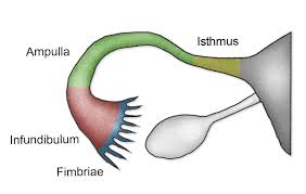
OVARIES

The ovaries are components of the female reproductive system and the endocrine system.
The female gonads are called the ovaries. In this article, we will initially look at the basic function, location, components and clinical significance of the ovaries. The latter part of the article will cover the ligaments associated with the ovaries and their vasculature, lymphatic drainage and innervation.
In both the males and females, the gonads develop within the mesonephric ridge and descend through the abdomen. However, unlike the testes, the ovaries stop in the pelvis.
The ovaries are paired, oval organs attached to the posterior surface of the broad ligament of the uterus by the mesovarium (a fold of peritoneum, continuous with the outer surface of the ovaries).
Each ovary is whitish in color and located alongside the lateral wall of the uterus in a region called the ovarian fossa. The ovarian fossa is the region that is bounded by the external iliac artery and in front of the ureter and the internal iliac artery. This area is about 4 cm x 3 cm x 2 cm in size.[3][4]
Function:
The ovaries produce oocytes and the hormones, oestrogen and progesterone.
At puberty, the ovary begins to secrete increasing levels of hormones. Secondary sex characteristics begin to develop in response to the hormones. The ovary changes structure and function beginning at puberty.[2] Since the ovaries are able to regulate hormones, they also play an important role in pregnancy and fertility. When egg cells (oocytes) are released from the fallopian tube, a variety of feedback mechanisms stimulate the endocrine system, which cause hormone levels to change.[10] These feedback mechanisms are controlled by the hypothalamus and pituitary glands. Messages or signals from the hypothalamus are sent to the pituitary gland. In turn, the pituitary gland releases hormones to the ovaries. From this signaling, the ovaries release their own hormones.
Position:
The ovaries are attached to the back of the broad ligaments within the peritoneal cavity.
Relations:
* Anterior to the ovaries are the broad ligaments.
* Posterior to the ovaries are the intestines.
* Lateral to the ovaries are the infundibulopelvic ligaments and the side walls of the pelvis.
* Superior to the ovaries lie the uterine tubes.
* Medial to the ovaries lie the ovarian ligaments and the uterus.
Supports:
The ovary is attached to the broad ligament but is supported from above by the ovarian ligament medially and the infundibulopelvic ligament laterally.
Structure:
The ovary is composed of a medulla and cortex, covered It with germinal epithelium.
* The medulla is the supporting framework, which is made of fibrous tissue; the ovarian blood vessels,lymphatics and nerves travel through it. The hilum where these vessels enter lies just where the ovary attached to the broad ligament and this area is called the mesovarium The cortex is the functioning part of the ovary contains the ovarian follicles in different stages of development, surrounded by stroma. The outer layer is formed of fibrous tissue known as the tunica albuginea. Over this lies the germinal epithelium which is a modification of the peritoneum.
Components of the Ovary:
The ovary has three main histological features:
- Surface – formed by simple cuboidal epithelium (known as germinal epithelium). Underlying this layer is a dense connective tissue capsule.
- Cortex – comprised of a connective tissue stroma and numerous ovarian follicles. Each follicle contains an oocyte, surrounded by a single layer of follicular cells.
- Medulla – formed by loose connective tissue and a rich neurovascular network, which enters via the hilum of the ovary.
Blood Supply:
Blood is supplied to the ovaries from the ovarian arteries and drains via the ovarian veins. The right ovarian vein joins the inferior vena cava, but the left returns its blood to the left renal vein.
Lymphatic Drainage:
Lymphatic drainage is to the lumbar glands.
Nerve Supply:
The nerve supply is from the ovarian plexus.