Fallopian Tubes

The uterine tubes, also known as fallopian tubes, oviducts and salpinges, are two very fine tubes leading from the ovaries into the uterus.
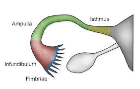
Each tube is a muscular hollow organ[3] that is on average between 10 and 14 cm (3.9 and 5.5 in) in length, with an external diameter of 1 cm (0.39 in).[4] It has four described parts: the intramural part, isthmus, ampulla, and infundibulum with associated fimbriae. Each tube has two openings: a proximal opening nearest to the uterus, and a distal opening nearest to the ovary. The fallopian tubes are held in place by the mesosalpinx, a part of the broad ligament mesentery that wraps around the tubes. Another part of the broad ligament, the mesovarium suspends the ovaries in place.[5]
The fallopian tube allows the passage of an egg from the ovary to the uterus. When an oocyte is developing in an ovary, it is surrounded by a spherical collection of cells known as an ovarian follicle. Just before ovulation, the primary oocyte completes meiosis I to form the first polar body and a secondary oocyte, which is arrested in metaphase of meiosis II.
Function:
The uterine tube propels the ovum towards the uterus, receives the spermatozoa as they travel upwards and provides a site for fertilization. It supplies the fertilized ovum with nutrition during its continued journey to the uterus.
Position:
The uterine tubes extend laterally from the cornua of the uterus towards the side walls of the pelvis. They arch over the ovaries, the fringed ends hovering near the ova- ries in order to receive the ovum.
Relations:
* Anterior, posterior and superior to the uterine tubes are the peritoneal cavity and the intestines.
* Lateral to the uterine tubes are the side walls of the pelvis.
* Inferior to the uterine tubes lie the broad ligaments and the ovaries.
* Medial to the two uterine tubes lies the uterus.
Supports:
The uterine tubes are held in place by their attachment to the uterus. The peritoneum folds over them, draping down below as the broad ligaments and extending at the sides to form the infundibulopelvic ligaments.
Structure:
Each tube is 10 cm long. The lumen of the tube provides an open pathway from the outside to the peritoneal cavity. The uterine tube has four portions • The interstitial portion is 1.25 cm long and lies within the wall of the uterus. Its lumen is 1 mm wide. The isthmus is another narrow part that extends for 2.5 cm from the uterus.
* The ampulla is the wider portion, where fertilization usually occurs. It is 5 cm long. • The infundibulum is the funnel-shaped fringed end that is composed of many processes known as fimbriae. One fimbria is elongated to form the ovarian fimbria, which is attached to the ovary.
Layers:
The lining of the uterine tubes is a mucous membrane of ciliated cubical epithelium that is thrown into complicated folds known as plicae. These folds slow the ovum down on its way to the uterus. In this lining are goblet cells that produce a secretion containing glycogen to nourish the oocyte.
Beneath the lining is a layer of vascular connective tissue.
The muscle coat consists of two layers: an inner circular layer and an outer longitudinal layer, both of smooth muscle. The peristaltic movement of the uterine tube is due to the action of these muscles.
The tube is covered with peritoneum but the infundibulum passes through it to open into the peritoneal cavity.
Blood Supply:
The blood supply is via the uterine and ovarian arteries, returning by the corresponding veins.
Lymphatic Drainage:
Lymph is drained to the lumbar glands.
Nerve Supply:
The nerve supply is from the ovarian plexus.